

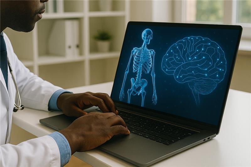
At WISE, we support innovations in education that respond to the evolving needs of learners and educators. This article highlights how immersive technologies are transforming medical education in practice at Weill Cornell Medicine–Qatar (WCM-Q), demonstrating the potential of 3D simulations and virtual reality to deepen anatomical understanding, enhance accessibility, and better prepare future physicians for modern clinical environments.
Introduction
Anatomy education has long been a foundation of medical training, evolving significantly over the centuries. This journey spans from traditional cadaveric dissection—the gold standard of anatomical education—to today’s digital transformation. While historical methods relied heavily on textbooks, 2D illustrations, and physical models, they served as the foundation for understanding human structure. These conventional approaches, though fundamental, had natural limitations in their ability to demonstrate the dynamic nature of anatomical relationships.
The late 20th century saw the integration of multimedia tools, with PowerPoint presentations and educational videos supplementing traditional teaching methods. However, these digital additions, while expanding the educational toolkit, still constrained learners to passive observation rather than active exploration. The inability to manipulate, layer, and interact with anatomical structures in real-time remained a significant teaching challenge, highlighting the need for more sophisticated educational technologies that could bridge the gap between theoretical knowledge and practical understanding.
The Use of Technology at Weill Cornell Medicine-Qatar (WCM-Q)
The WCM-Q journey with integrating 3D and immersive technologies into anatomy education began about eight years ago, marking a significant evolution in how we approach anatomical teaching. While maintaining our commitment to traditional teaching methods, we strategically incorporated the Anatomage virtual dissection table and virtual reality (VR) technology, creating a dynamic and interactive learning environment that enhances traditional approaches and empowers students to explore anatomy in innovative ways.
The Anatomage Table
This is an interactive 3D anatomy visualization system, which has proven to be very useful for demonstrating complex anatomical relationships that are challenging to visualize in traditional dissection. It provides an unrestricted view of anatomical structures, allowing students to manipulate digital cadavers, isolate specific systems, and simulate dissections. Students can explore detailed, high-resolution 3D models of the human body, and visualize relationships between organs, muscles, nerves, and vasculature with precision.
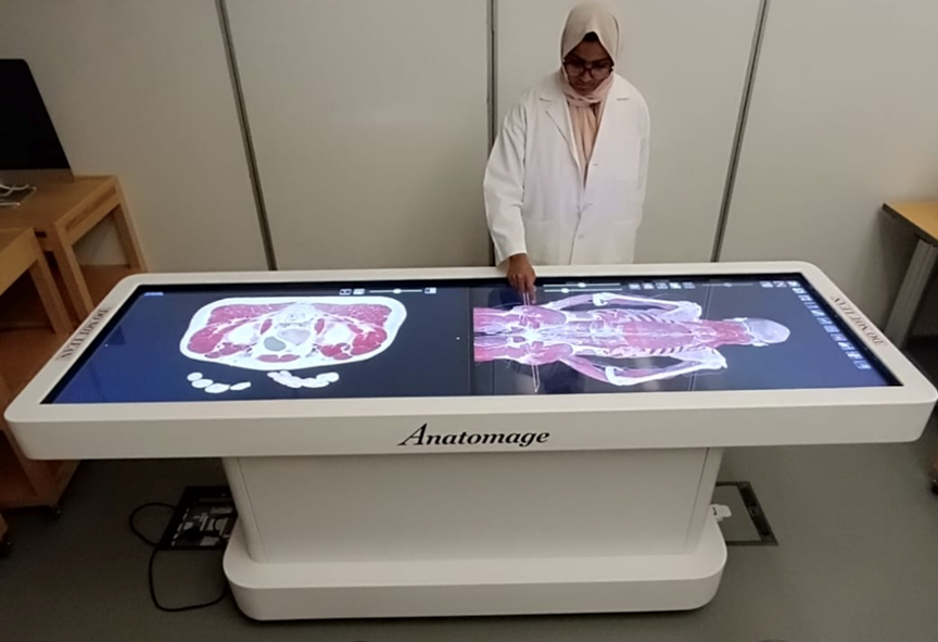
For example, students can isolate the brain, rotate it in 3D space, and virtually “dissect” layers to reveal deep structures like the basal ganglia, internal capsule, and ventricular system. The table also features unique capabilities like simulating cardiac conduction systems and the stages of childbirth. Its unlimited accessibility has fostered independent learning, allowing students to review structures repeatedly at their own pace. This flexibility empowers students to revisit challenging concepts and bridge the gap between theoretical knowledge and practical application.
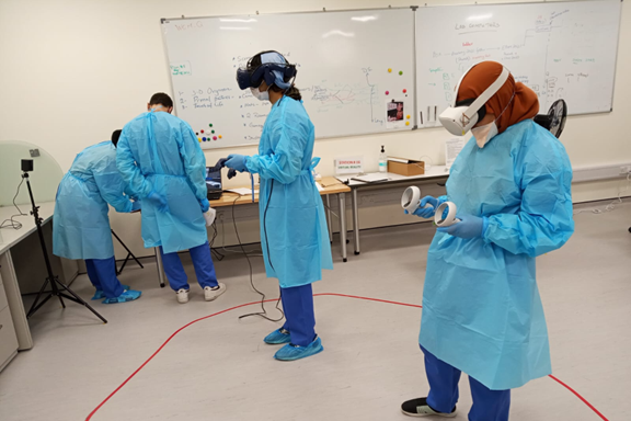
VR Technology
Along with the Anatomage table, we utilize VR technology to create an immersive and interactive learning environment, offering students an effective way to understand complex three-dimensional structures. Through VR headsets and anatomy software, students can explore complex regions like the perineum and pelvis, where understanding spatial relationships is traditionally challenging. They can visualize the layered arrangement of the pelvic diaphragm, the spatial relationships between pelvic organs, and the courses of neurovascular structures.
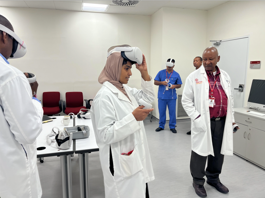
The ability to systematically remove layers, rotate structures, and isolate specific components has significantly enhanced students’ grasp of complex anatomical concepts. The portability of VR technology has extended learning beyond the classroom, enabling students to access anatomical materials and join virtual sessions both on campus and at home, maintaining interactive learning experiences despite physical distance. This capability has opened new possibilities for reaching students across different locations and accommodating diverse learning schedules.
By actively engaging with 3D models in a collaborative VR environment, students gain a deeper understanding of spatial relationships and reinforce memory retention. When integrated with hands-on anatomy sessions, this immersive experience bridges the gap between theory and practice, making learning more intuitive and impactful.
Student feedback on these technological innovations has been positive, particularly regarding their ability to grasp complex anatomical concepts and build confidence in structure identification. These digital tools have been very useful for self-directed learning and unlimited content review. The clean, controlled environment of virtual learning has made anatomy education more accessible to students with diverse learning styles and sensory sensitivities. Our experience has shown that when integrated with traditional teaching methods, these technologies create a comprehensive learning experience that enhances both competence and confidence across our diverse student population.
Challenges/Limitations
While the Anatomage virtual dissection table and VR systems have revolutionized anatomy education, they are not without limitations. One significant challenge is the inability of these technologies to foster empathy and humanity, which are integral to traditional cadaver dissection. Confronting the reality of death and understanding human mortality are experiences that virtual tools cannot replicate. Additionally, most software relies on standardized anatomical models, which lack the full complexity of individual variations in muscles, bones, tendons, and other structures.
Technical and logistical hurdles also arise when implementing these technologies. Poorly designed software, network issues, and the need for skilled operators can hinder seamless integration into the curriculum. Furthermore, overreliance on virtual tools risks diminishing students’ engagement with physical anatomy, potentially leading to gaps in understanding real-world variability, such as intricate differences in muscles, veins, or pathologies.
Future Directions
Looking ahead, we anticipate several exciting advancements in our anatomy unit. We are currently exploring Medical Holodeck technology to create customized anatomical images, marking a significant step toward tailoring digital resources to meet specific educational goals. As these technologies evolve, we remain committed to evaluating their educational impact through research and assessment. This ensures that each technological innovation genuinely enhances learning outcomes and equips our students for the increasingly digital future of modern medicine.